
Effects of Carrot (Daucus carota) Stalk on Blood Glucose Level, Biochemical Functions and Serum Antioxidant Activity in Alloxan-Induced Diabetic Rats
-
Ikechukwu Johnlouis Obidike


Department of Biochemistry, Michael Okpara University of Agriculture, Umudike, Abia, Nigeria
Mbah Esther NgoziDepartment of Biochemistry, Michael Okpara University of Agriculture, Umudike, Abia, Nigeria
| Received 25 Oct, 2023 |
Accepted 29 Jan, 2024 |
Published 20 Feb, 2024 |
Background and Objective: Carrot (Daucus carota) is a root vegetable and its stalk which is the subject of this study is discarded as waste. The aim of this study is to determine the effects of carrot (Daucus carota) stalk on blood glucose level, biochemical functions, liver histo-morphology and serum antioxidant activity in alloxan-induced diabetic rats. Materials and Methods: Following the induction of diabetes using alloxan, the animal were treated with 400 and 800 mg/kg of the extract and glibenclamide, a standard drug for 21 days. The rats’ biochemical parameter, liver histo-morphology and serum antioxidant activities were examined using standard procedures and analysed by one-way ANOVA using SPSS version 20. The effect of Daucus carotastalk extract on the blood glucose level of the diabetic rats compared favourably with that of glibenclamide from day 0 to 21. Results: The results of the biochemical assays show significant (p<0.05) reduction in liver enzymes (AST) -11.11 and -15.69% relative to alloxan group and -3.6% relative to glibenclamide; while ALT showed -6.82 and -2.38% change relative to alloxan and glibenclamide, respectively. This same pattern was also observed in total bilirubin concentration and also extended to the calculated ratios (AST:ALT, ALT:ALP and AST:ALP) including the serum antioxidant activity. The liver histo-morphological presentation seemingly supports the biochemical parameter results showing possible ameliorative effects. Conclusion: Conclusively, it could be observed that the extract of Daucus carota stalk has antidiabetic properties which are comparable to that of glibenclamide and ameliorated the effects of the hepatic injury caused by alloxan in diabetic rats.
INTRODUCTION
Carrot (Daucus carota); a versatile vegetable that appears in several colors, shapes and sizes1 is one of the most important root vegetable plants in the world and can be eaten both raw and cooked2. The quality of fruits and vegetables is determined by nutritional value and carrots have been ranked 10th among fruit and vegetables3. Carrots contain molybdenum; a rare trace element rarely found in many vegetables4 and has been also found to be a good source of dietary and high concentration of carotenoids, a family of antioxidants which mops out free radicals implicated in conditions such as DNA damage, cellular membrane damage and diabetes mellitus5.
Diabetes mellitus (DM) is a metabolic disease characterized by elevated blood glucose. Type 1 diabetes is caused by insufficient insulin production by the pancreas, while type 2 diabetes is caused by impaired insulin resistance (IR) of the liver and skeletal muscle, two responsible tissues, to available insulin6. Hyperglycemia is the overall result, which can cause fatal complications as well as serious health issues. Approximately 387 million people worldwide suffer from diabetes mellitus (DM), with a prevalence rate of 8.3%. Many of these individuals, particularly in Africa, go undiagnosed7,8. About 2.7% of all deaths are caused by diabetes mellitus (DM), which is also linked to ischemic heart disease and stroke9, the top two causes of death globally10. Diabetes is becoming more commonplace, mainly in low- and middle-income countries worldwide11. It is closely associated with obesity and sedentary lifestyles. Alloxan is a potent agent for inducing experimental diabetes mellitus12, particularly type I diabetes mellitus13. A urea derivative called alloxan selectively necrotizes the β-cells in the pancreatic islets. Therefore, by adjusting the dosage of alloxan used, it has been widely used to induce experimental diabetes in animals such as rabbits, rats, mice and dogs with varying degrees of disease severity14. Owing to alloxan’s structural resemblance to glucose, the mechanism responsible for alloxan-induced diabetes is selective uptake of the compound15.
Consumption of Daucus carota stalk, a waste product of a conventionally available fruit with high antioxidant potential could improve the management of diabetes mellitus and redirect the management of the condition from synthetic drugs which are widely believed to have a long-term adverse effect to a more natural management routine believed to be safer and promote environmental sanitation when the waste is channelled into pharmacological production hence this study aimed at determining the effects of carrot (Daucus carota) stalk on biochemical functions and histo-morphology of the liver in alloxan-induced diabetic rats.
MATERIALS AND METHODS
Collection and identification of sample: The study took place in the laboratory unit of Michael Okpara University of Agriculture, Umudike Abia State, Nigeria between March, 2022 to August, 2022. Carrot stalks were bought from Eke Ukwu Owerri Imo State Nigeria. The stalk was identified as Daucus carota stalk by a taxonomist Prof. G.G.E. Osuagwu in the Department of Plant Science and Biotechnology, Michael Okpara University of Agriculture Umudike, Abia State, Nigeria.
Preparation of extract: The stalks were thoroughly washed with tap water to remove sand and other debris that may contaminate the sample. The washed samples were then air-dried under shade on a laboratory bench for six weeks. Thereafter the dried plant samples were pulverized using Corona manual blender Model 0719, manufactured in Germany. Forty grams of the powdered sample were extracted with 96% ethanol in a Soxhlet extractor (model SE-6P Infitek, China). The temperature was maintained at 60°C throughout the 48 hrs extraction period. At the end of the period the extract was dried in a hot air oven (IEC60068-2-2 Dongguan China) maintained at 40°C to obtain a dark green semi-solid sample which weighed 2.00 g. The crude ethanol extract was kept in an air-tight container and stored in a refrigerator (Thermocool, Nigeria) at 10°C until the time of use. Percentage yield of the extract was calculated using the formula16:
Where:
X |
= |
Weight of dried Daucus carota stalk extract after extraction (2.00 g) |
|
Q |
= |
Weight of Daucus carota stalk extract powdered plant material before extraction |
Study design for the biochemical estimation of Daucus carota stalk extract: Thirty male albino rats were randomly divided into six groups of six rats in each group and were in addition to feed and water assigned treatments. They were acclimatized for 12 before the random division into in an aluminum cage. All of the animals were kept in standard laboratory settings, with 12 hrs light and dark cycles and room temperature. They also had unlimited access to food and water. Every rule pertaining to the use and maintenance of lab animals was scrupulously followed.
The animals were grouped as such:
Group A |
: |
Feed and water |
|
Group B |
: |
Received 400 mg/kg of Daucus carota stalk extract |
|
Group C |
: |
Untreated diabetic rats (alloxan160 mg/kg) |
|
Group D |
: |
Treated diabetic rats (alloxan 160+400 mg/kg of Daucus carota stalk extract) |
|
Group E |
: |
Treated diabetic rats (alloxan 160+800mg/kg of Daucus carota stalk extract) |
|
Group F |
: |
Treated diabetic rats (alloxan 160+3 mg/kg glibenclamide-a standard anti-diabetic drug) |
Induction of diabetes mellitus: Twenty rats were administered alloxan monohydrate (Sigma Chemical Co. USA) dissolved in 10% (w/v) normal saline at a dose of 160 mg/kg body weight by a single intraperitoneal cavity injection (hind limb), while the remaining ten rats were non-diabetic and served as control. The alloxan used was prepared by dissolving 0.25 g of alloxan in 3 mL of distilled water. After 5 days of alloxan administration, blood was obtained by tail snip from each rat and tested for glucose level to confirm the development of diabetes using a glucose meter (Roche Co. Germany). Rats with fasting glucose levels above 190 mg/dl were considered diabetic and hence were used in carrying out this research work. All guidelines involving the use and care of laboratory animals were duly observed.
Tissue preparation: The experimental animals were humanely sacrificed at the end of the study. Gross lesions were recorded as observed during the post-mortem examination. Sections of the pancreas, liver and kidney were collected for histopathological examination. The collected sample from each of the six groups was fixed in 10% phosphate-buffered formalin for a minimum of 48 hrs. The tissues were subsequently trimmed, dehydrated in 4 grades of alcohol (70, 80, 90 and absolute alcohol), cleared in 3 grades of xylene and embedded in molten wax. On solidifying, the blocks were sectioned, 5 μm thick with a rotary microtome, floated in water bathing and incubated at 60°C for 30 min. The 5 μm thick sectioned tissues were subsequently cleared in 3 grades of xylene and rehydrated in 3 grades of alcohol (90, 80 and 70%). The sections were then stained with hematoxylin for 15 min. Blueing was done with ammonium chloride. Differentiation was done with 1% acid alcohol before counterstaining with eosin. Permanent mounts were made on degreased glass slides using a mountant, DPX.
Determination of Aspartate Aminotransferase (AST) and Alanine Aminotransferase (ALT): The determination of Aspartate Aminotransferase (AST) and Alanine Aminotransferase (ALT) in serum was done according to the method described by Johnlouis et al.16.
Determination of Alkaline Phosphatase (ALP): The determination of alkaline phosphatase in whole blood was done according to the method described by Johnlouis et al.16.
Bilirubin estimation: The serum total bilirubin in the serum was estimated by the endpoint colorimetric method as described by Johnlouis et al.16. This is based on the principle that bilirubin reacts with diazotized sulfanilic acid to form zero azo compound producing a colour-measured photometrically at 555 nm.
Determination of serum total protein: The serum total protein was estimated by the biuret method described by Johnlouis et al.16. This is based on the process by which the biuret reagent's cupric acid ion interacts with protein peptide bonds to form a violet color in an alkaline medium. The amount of color that forms is proportional to the total amount of protein in the sample and is measured photometrically at 540 nm.
Estimation of superoxide dismutase (SOD): The method of autooxidation by pyrogallol was used by Egbuonu et al.17. Pyrogallol autooxides rapidly in an alkaline solution generating superoxide ions. The SOD inhibits its auto-oxidation dismutating the superoxide ions to hydrogen peroxide and molecular oxygen. The activity of 50% inhibition by SOD was measured at 450 nm.
Determination of serum catalase: The Egbuonu et al.17 method was utilized to determine the catalase activity. The idea behind this is that hydrogen peroxide can be split by catalase. The addition of the dichromate/acetic acid reagent stopped the reaction after a predetermined amount of time. Dichromate in acetic acid was reduced to chromic acetate and an unstable perchromic intermediate when heated in the presence of hydrogen peroxide. By measuring the amount of chromic acetate produced at 610 nm, the amount of hydrogen peroxide that was still present was ascertained.
Determination of reduced glutathione: Reduced glutathione was estimated using the Egbuonu et al.17 method. It entails giving compounds with sulfhydryl groups a yellow color by reacting them with the Ellman’s reagent (5,5 Dithiobis(2-Nitrobenzoic Acid) (DTNB). At 412 nm, the absorbance is measured. Estimation of extent of lipid peroxidation (malondialdehyde).
According to Wallin et al.18, spectrophotometric measurement of the lipid peroxidation product, malondialdehyde (MDA), was used to estimate lipid peroxidation. When lipids break down, compounds like pentane, ethane and malondialdehyde which are formed from the n-terminal carbons of three and six fatty acids, respectively and fatty acids with two or more double bonds are produced. Thiobarbituric acid and MDA combine to generate a reddish-pink complex that has a maximum absorption at 532 nm in an acidic solution.
Statistical analysis: Data were analyzed by One-way Analysis of Variance (ANOVA) using Statistical Package of Social Sciences (SPSS) software version 20.0 for windows. Results were expressed as Mean±Standard Error (SE) and difference in mean tested using post hoc LSD. A level of p<0.05 was considered significant.
RESULTS
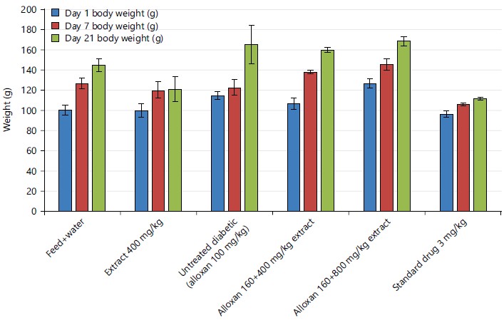
The effects of ethanol extract of Daucus carota stalk on body weight in alloxan induced diabetic rats is shown in Fig. 1. The result shows a significant increase in the body weight of animals across all the groups from day 1 to 21. Group 1 weighed 100.54 g on day 1, 126.73 g on day 7 and 144.96 g on day 21. Group 2 weighed 99.97 g on day 1, 120.07 g on day 7 and 120.95 g on day 21. Group 3 weighed 114.63 g on day 1, 122.88 g on day 7 and 165.45 g on day 21. Group 4 weighed 106.75 g on day 1, 137.81 g on day 7 and 159.68 g on day 21. Group 5 weighed 126.45 g on day 1, 145.47 g on day 7 and 168.54 g on day 21. Group 6 weighed 96.36 g on day 1, 105.86 g on day 7 and 111.72 g on day 21.
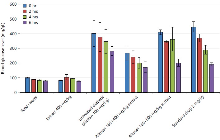
Figure 2 shows the effects of ethanol extract of Daucus carota stalk on blood glucose levels in alloxan induced diabetic rats. There was an increase in blood glucose across the treated groups at 0 hr as expected. However, a significant decrease was noticed across the groups co-treated Daucus carota stalk extract and glibenclamide group with increase in time.
|
|
Table 1 shows the effects of ethanol extract of Daucus carota stalk on AST activity in alloxan induced diabetic rats. Serum Aspartate Transaminase (AST) level was significantly (p<0.05) elevated in Group C when compared to other groups. However, the highest reduction in activity was noticed in the group that received 800 mg/kg as shown by percentrage of change relative to glibenclamide (-3.62%).
Table 2 shows the effects of ethanol extract of Daucus carota stalk on ALT activity in alloxan-induced diabetic rats. The lowest activity was seen in the low-dose cotreated group even lower than the standard drug as indicated by the percentage of change relative to glibenclamide (-2.38%).
| Table 1: | Effects of ethanol extract of Daucus carota stalk on AST activity in alloxan-induced diabetic rats | |||
| Group | AST |
Change relative to alloxan (%) |
Change relative to glibenclamide (%) |
| A = Feed+water | 28.67±0.76 |
-25.05 |
-14.32 |
| B = Extract group (400 mg/kg b.wt., extract) | 34.83±1.38 |
-8.94 |
4.09 |
| C = Alloxan group (160 mg/kg b.wt.) | 38.25±2.66 |
0 |
14.31 |
| D = Extract cotreated group (alloxan 160+400 mg/kg of Daucus carota stalk) | 34.00±0.71 |
-11.11 |
1.61 |
| E = Extract cotreated group (alloxan 160+800 mg kg of Daucus carota stalk) | 33.25±0.48 |
-15.69 |
-3.62 |
| F = Extract cotreated group (alloxan 160+3 mg kg of glibenclamide) | 33.46±0.74 |
-12.52 |
0 |
| Values are Mean±SEM for n = 4, Difference is considered statistically significant at p<0.05, +: Higher and -: Lower | |||
| Table 2: | Effects of ethanol extract of Daucus carota stalk on ALT activity in alloxan induced diabetic rats | |||
| Group | ALT |
Change relative to alloxan (%) |
Change relative to glibenclamide (%) |
| A = Feed+water | 10.50±0.40 |
-4.55 |
0 |
| B = Extract group (400 mg/kg b.wt., extract) | 10.83±0.31 |
-1.55 |
3.14 |
| C = Alloxan group (160 mg/kg b.wt.) | 11.00±0.14 |
0 |
4.76 |
| D = Extract cotreated group (alloxan 160+400 mg/kg of Daucus carota stalk) | 10.25±0.63 |
-6.82 |
-2.38 |
| E = Extract cotreated group (alloxan 160+800 mg/kg of Daucus carota stalk) | 11.00±0.41 |
0 |
4.76 |
| F = Extract cotreated group (alloxan 160+3 mg/kg of glibenclamide) | 10.50±0.29 |
-4.55 |
0 |
| Values are Mean±SEM for n = 4, Difference is considered statistically significant at p<0.05, +: Higher and -: Lower | |||
| Table 3: | Effects of ethanol extract of Daucus carota stalk on ALP activity in alloxan induced diabetic rats | |||
| Group | ALP |
Change relative to alloxan (%) |
Change relative to glibenclamide (%) |
| A = Feed+water | 11.13±0.34 |
5.5 |
2.77 |
| B = Extract group (400 mg/kg b.wt., extract) | 11.90±0.17 |
12.8 |
-9.88 |
| C = Alloxan group (160 mg/kg b.wt.) | 10.95±0.37 |
0 |
-2.53 |
| D = Extract cotreated group (alloxan 160+400 mg/kg of Daucus carota stalk) | 10.90±0.38 |
3.32 |
0.65 |
| E = Extract cotreated group (alloxan 160+800 mg/kg of Daucus carota stalk) | 10.98±0.17 |
4.08 |
1.39 |
| F = Extract cotreated group (alloxan 160+3 mg/kg of glibenclamide) | 10.83±0.15 |
2.65 |
0 |
| Values are Mean±SEM for n = 4, Difference is considered statistically significant at p<0.05, +: Higher and -: Lower | |||
| Table 4: | Effects of ethanol extract of Daucus carota stalk on total bilirubin concentration in alloxan induced diabetic rats | |||
| Group | Total billirubin |
Change relative to alloxan (%) |
Change relative to glibenclamide (%) |
| A = Feed+water | 1.42±0.10 |
-18.39 |
5.18 |
| B = Extract group (400 mg/kg b.wt., extract) | 1.66±0.06 |
-4.6 |
22.96 |
| C = Alloxan group (160 mg/kg b.wt.) | 1.74±0.04 |
0 |
28.89 |
| D = Extract cotreated group (alloxan 160+400 mg/kg of Daucus carota stalk) | 1.14±0.04 |
-34.48 |
-15.56 |
| E = Extract cotreated group (alloxan 160+800 mg/kg of Daucus carota stalk) | 1.26±0.06 |
-27.58 |
-6.67 |
| F = Extract cotreated group (alloxan 160+3 mg/kg of glibenclamide) | 1.42±0.10 |
-22.41 |
0 |
| Values are Mean±SEM for n = 4, Difference is considered statistically significant at p<0.05, +: Higher and -: Lower | |||
Table 3 shows the effects of ethanol extract of Daucus carota stalk on ALP activity in alloxan induced diabetic rats. Alkaline Phosphatase (ALP) level was high in Group B compared to other groups. Compared to Group C (10.95±0.37), ALP level in Group A (11.13±0.34), was significantly (p<0.05) increased. Group D, E and F showed no statistical significance.
Table 4 shows the effects of ethanol extract of Daucus carota stalk on total bilirubin in alloxan-induced diabetic rats. The results showed reduced serum concentrations in the low dose co-treated group (400 mg/kg) as compared to the standard drug (glibenclamide).
Table 5 shows the effects of ethanol extract of Daucus carota stalk on total protein concentration in alloxan-induced diabetic rats. The results showed reduced serum concentrations in the low dose co-treated group (400 mg/kg) as compared to the standard drug (glibenclamide).
Table 6 shows the AST:ALT (ALT:AST) ratio of the ethanol extract of Daucus carota stalk on AST:ALT ratio in alloxan-induced diabetic rats. The result indicates lowest change relative percentage to alloxan in the normal group [-21.55 (27.59)] followed by the high dose [-15.80 (17.24)] while the highest was the low dose [-4.60 (3.45)].
| Table 5: | Effects of ethanol extract of Daucus carota stalk on total protein concentration in alloxan induced diabetic rats | |||
| Group | Total protein |
Change relative to alloxan (%) |
Change relative to glibenclamide (%) |
| A = Feed+water | 6.40±0.21 |
29.82 |
4.92 |
| B = Extract group (400 mg/kg b.wt., extract) | 5.78±0.15 |
17.24 |
-5.25 |
| C = Alloxan group (160 mg/kg b.w.t) | 4.93±0.19 |
0 |
-19.18 |
| D = Extract cotreated group (alloxan 160+400 mg/kg of Daucus carota stalk) | 6.05±0.18 |
22.72 |
-0.81 |
| E = Extract cotreated group (alloxan 160+800 mg/kg of Daucus carota stalk) | 5.95±0.17 |
20.69 |
2.46 |
| F = Extract cotreated group (alloxan 160+3 mg/kg of glibenclamide) | 6.10±0.13 |
23.73 |
0 |
| Values are Mean±SEM for n = 4, Difference is considered statistically significant at p<0.05, +: Higher and -: Lower | |||
| Table 6: | Effects of ethanol extract of Daucus carota stalk on AST:ALT ratio in alloxan induced diabetic rats | |||
| Group | AST:ALT (ALT:AST) |
Change relative to alloxan (%) |
Change relative to glibenclamide (%) |
| A = Feed+water | 2.73 (0.37) |
-21.55 (27.59) |
-14.42 (19.35) |
| B = Extract group (400 mg/kg b.wt., extract) | 3.22 (0.31) |
-7.47 (6.90) |
0.94 (0.00) |
| C = Alloxan group (160 mg/kg b.wt.) | 3.48 (0.29) |
0.00 (0.00) |
8.78 (-6.45) |
| D = Extract cotreated group (alloxan 160+400 mg/kg of Daucus carota stalk) | 3.32 (0.03) |
-4.60 (3.45) |
-4.08 (-3.23) |
| E = Extract cotreated group (alloxan 160+800 mg/kg of Daucus carota stalk) | 2.93 (0.34) |
-15.80 (17.24) |
-8.15 (9.67) |
| F = Extract cotreated group (alloxan 160+3 mg/kg of glibenclamide) | 3.19 (0.31) |
-8.33 (6.90) |
0.00 (0.00) |
| Values are Mean±SEM for n = 4, Difference is considered statistically significant at p<0.05, +: Higher and -: Lower | |||
| Table 7: | Effects of ethanol extract of Daucus carota stalk on AST:ALP ratio in alloxan induced diabetic rats | |||
| Group | AST:ALP (ALP:AST) |
Change relative to alloxan (%) |
Change relative to glibenclamide (%) |
| A = Feed+water | 2.57 (0.39) |
-29.20 (39.29) |
-16.83 (21.88) |
| B = Extract group (400 mg/kg b.wt., extract) | 2.89 (0.34) |
-20.39 (21.43) |
-6.47 (6.25) |
| C = Alloxan group (160 mg/kg b.wt.) | 3.36 (0.28) |
0.00 (0.00) |
17.48 (-12.50) |
| D = Extract cotreated group (alloxan 160+400 mg/kg of Daucus carota stalk) | 3.12 (0.32) |
14.05 (14.29) |
0.97 (0.00) |
| E = Extract cotreated group (alloxan 160+800 mg/kg of Daucus carota stalk) | 2.94 (0.34) |
-19.01 (21.43) |
-4.85 (6.25) |
| F = Extract cotreated group (alloxan 160+3 mg/kg of glibenclamide) | 3.09 (0.32) |
-14.88 (14.29) |
0.00 (0.00) |
| Values are Mean±SEM for n = 4, Difference is considered statistically significant at p<0.05, +: Higher and -: Lower | |||
| Table 8: | Effects of ethanol extract of Daucus carota stalk on ALT:ALP ratio in alloxan induced diabetic rats | |||
| Group | ALT:ALP (ALP:ALT) |
Change relative to alloxan (%) |
Change relative to glibenclamide (%) |
| A = Feed+water | 0.94 (1.06) |
-9.62 (10.42) |
-3.09 (2.91) |
| B = Extract group (400 mg/kg b.wt., extract) | 0.91 (1.10) |
-12.50 (14.58) |
-6.19 (6.80) |
| C = Alloxan group (160 mg/kg b.wt.) | 1.04 (0.96) |
0.00 (0.00) |
7.22 (-6.80) |
| D = Extract cotreated group (alloxan 160+400 mg/kg of Daucus carota stalk) | 0.94 (1.06) |
-9.62 (10.42) |
-3.09 (2.91) |
| E = Extract cotreated group (alloxan 160+800 mg/kg of Daucus carota stalk) | 1.00 (1.00) |
-3.84 (4.17) |
3.09 (-2.91) |
| F = Extract cotreated group (alloxan 160+3 mg/kg of glibenclamide) | 0.97 (1.03) |
-6.73 (7.29) |
0.00 (0.00) |
| Values are Mean±SEM for n = 4, Difference is considered statistically significant at p<0.05, +: Higher and -: Lower | |||
Table 7 shows the AST:ALP (ALP:AST) change relative to alloxan and glibenclamide. The result indicated that the change relative to alloxan was highest in the normal group [-29.20 (39.29)] while those of the co-treated groups were in dose-dependent manner {[14.05 (14.29)] for low dose and [-19.01 (21.43)] for high dose}.
Table 8 shows the ALT:ALP (ALP:ALT) ratio relative to alloxan and glibenclamide, respectively. Change relative to extract group showed the highest change [-12.50 (14.58)] while the co-treated groups showed dose-dependent relative change {[-9.62 (10.42)] for low dose and [-3.84 (4.17)]} for high dose.
Table 9 shows the effects of ethanol extract of Daucus carota stalk on in vivo antioxidant assay (GSH) in alloxan-induced diabetic rats. The results show reduced antioxidant figures in the extract co-treated groups when compared to the glibenclamide group as indicated by a negative percentage change relative to glibenclamide.
| Table 9: | Effects of ethanol extract of Daucus carota stalk on GSH concentration in alloxan induced diabetic rats | |||
| Group | GSH (mg/dl) |
Change relative to control (%) |
Change relative to glibenclamide (%) |
| A = Feed+water | 2.52±0.08 |
9.57 |
-13.99 |
| B = Extract group (400 mg/kg b.wt., extract) | 2.35±0.13 |
2.17 |
-19.8 |
| C = Alloxan group (160 mg/kg b.wt.) | 2.30±0.04 |
0 |
-21.5 |
| D = Extract cotreated group (alloxan 160+400 mg/kg of Daucus carota stalk) | 2.63±0.11 |
14.35 |
-10.24 |
| E = Extract cotreated group (alloxan 160+800 mg/kg of Daucus carota stalk) | 2.75±0.06 |
19.57 |
-6.14 |
| F = Extract cotreated group (alloxan 160+3 mg/kg of glibenclamide) | 2.93±0.05 |
27.39 |
0 |
| Values are Mean±SEM for n = 4, Difference is considered statistically significant at p<0.05, +: Higher and -: Lower | |||
| Table 10: | Effects of ethanol extract of Daucus carota stalk on superoxide dismutase activity in alloxan induced diabetic rats | |||
| Group | SOD (IU/L) |
Change relative to control (%) |
Change relative to glibenclamide (%) |
| A = Feed+water | 1.09±0.01 |
-0.91 |
-8.4 |
| B = Extract group (400 mg/kg b.wt., extract) | 1.08±0.01 |
-1.82 |
-9.24 |
| C = Alloxan group (160 mg/kg b.wt.) | 1.10±0.01 |
0 |
-7.56 |
| D = Extract cotreated group (alloxan 160+400 mg/kg of Daucus carota stalk) | 1.13±0.01 |
2.73 |
-5.04 |
| E = Extract cotreated group (alloxan 160+800 mg/kg of Daucus carota stalk) | 1.15±0.01 |
4.55 |
-3.36 |
| F = Extract cotreated group (alloxan 160+3 mg/kg of glibenclamide) | 1.19±0.00 |
8.18 |
0 |
| Values are Mean±SEM for n = 4, Difference is considered statistically significant at p<0.05, +: Higher and -: Lower | |||
| Table 11: | Effects of ethanol extract of Daucus carota stalk on total protein concentration in alloxan induced diabetic rats | |||
| Group | CAT (IU/L) |
Change relative to alloxan (%) |
Change relative to glibenclamide (%) |
| A = Feed+water | 1.33±0.02 |
31.68 |
12.71 |
| B = Extract group (400 mg/kg b.wt., extract) | 1.22±0.06 |
20.79 |
3.39 |
| C = Alloxan group (160 mg/kg b.wt.) | 1.01±0.09 |
0 |
-15.41 |
| D = Extract cotreated group (alloxan 160+400 mg/kg of Daucus carota stalk) | 1.15±0.02 |
13.84 |
-2.54 |
| E = Extract cotreated group (alloxan 160+800 mg/kg of Daucus carota stalk) | 1.15±0.01 |
13.84 |
-2.54 |
| F = Extract cotreated group (alloxan 160+3 mg/kg of glibenclamide) | 1.18±0.01 |
16.83 |
0 |
| Values are Mean±SEM for n = 4, Difference is considered statistically significant at p<0.05, +: Higher and -: Lower | |||
| Table 12: | Effects of ethanol extract of Daucus carota stalk on malondialdehyde (MDA) concentration in alloxan induced diabetic rats | |||
| Group | MDA (mg/mL) |
Change relative to alloxan (%) |
Change relative to glibenclamide (%) |
| A = Feed+water | 5.58±0.08 |
-12.13 |
9.2 |
| B = Extract group (400 mg/kg b.wt., extract) | 5.97±0.21 |
-5.98 |
16.83 |
| C = Alloxan group (160 mg/kg b.wt.) | 6.35±0.21 |
0 |
24.27 |
| D = Extract cotreated group (alloxan 160+400 mg/kg of Daucus carota stalk) | 5.38±0.17 |
-15.28 |
5.28 |
| E = Extract cotreated group (alloxan 160+800 mg/kg of Daucus carota stalk) | 5.13±0.02 |
-19.21 |
16.83 |
| F = Extract cotreated group (alloxan 160+3 mg/kg of glibenclamide) | 5.11±0.10 |
-19.53 |
0 |
| Values are Mean±SEM for n = 4, Difference is considered statistically significant at p<0.05, +: Higher and -: Lower | |||
The result for the SOD activity (Table 10) showed a dose-dependent increase in the SOD activity compared to that of the alloxan group. This is further buttressed by a dose-dependent increase in the percentage change relative to alloxan group (2.73) for low doses and (4.55) for high doses.
The result for CAT activity (Table 11) showed increased CAT activity in the co-treated groups compared to the alloxan group. The change relative to alloxan group also indicated a positive change.
The MDA (Table 12) in the co-treated groups were all within a comparable range as compared to the glibenclamide and were all lower than the untreated group. The calculation for change relative to alloxan showed a dose-dependent decrease (-15.28) for low doses and (-19.21) for high doses.
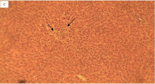
Photomicrograph plates: The liver section (Fig. 3) of the control group showed random, multifocal areas of necrosis (arrow) with no infiltration of inflammatory leucocytes.
|
|
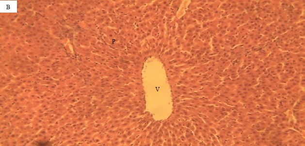
Section of the liver for the extract group (Fig. 4) showed no periportal vacuolar degeneration of the hepatocytes. This show clear vacuoles which do not displace the nucleus in their cytoplasm central vein (V) and portal area (P).
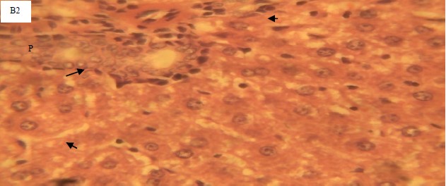
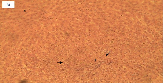
The liver section of the diabetic group (Fig. 5) showed a mild vacuolar degeneration of the hepatocytes. Affected hepatocytes appear swollen and show multiple coalescent clear vacuoles which displaced the nucleus in their cytoplasm.
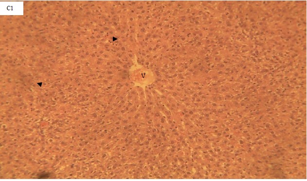
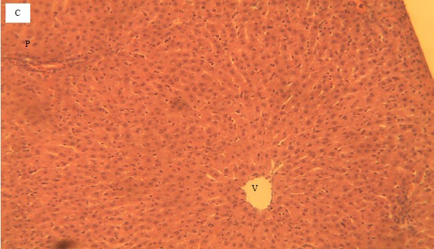
The liver section of the animals co-treated alloxan with 400 mg/kg (Fig. 6) showed the normal hepatic histo-architecture.
The liver section of animals co-treated alloxan with 800 mg/kg Daucus caroto stalk (Fig. 7) showed cytoplasmic vacuoles which do not displace the nuclei.
|
|
|
|
The liver section of animals co-treated alloxan with 3 mg/kg standard drug (Fig. 8) did not show any significant histopathological lesion. Central vein (V), portal area (P), normal renal tubule (RT), H&E×100 and ×400.
DISCUSSION
The result indicated that Daucus carota stalk showed antihyperglycemic effects on aloxan-induced animals. It also showed reduced liver enzyme activities (AST, ALT and ALP) which is indicative of positive ameliorative effects on the hepatotoxicity induced by alloxan. This is further buttressed by the histomorphological result which showed improved liver architecture in the histographs of the groups co-treated different concentrations of Daucus carota stalk extract. The result also showed that the extract has antioxidant potential hence, improved serum antioxidant activities were seen in the cotreated groups as compared to the alloxan group.
Sustained hyperglycaemia is implicated as an indicator of the onset of diabetes mellitus19. Green leafy vegetables may enhance falls in body weights usually seen in diabetic animals20,21. The effect of the extract on the blood glucose level of the diabetic rats also compared favourably with that of glibenclamide, a standard anti-diabetic and hypoglycaemic agent used thus agreed with the report of Ranjbar et al.22. The observed hypoglycaemic property may be indicative that the extract may contain active compounds with hypoglycaemic properties and may have achieved this hypoglycaemic property through increased peripheral utilization or increased insulin secretion23, inhibition of endogenous gluconeogenesis or inhibition of intestinal glucose absorption24.
The major enzymes assayed for diagnosis of hepatic injuries include AST, ALT and ALP with ALT being implicated as the most specific for hepatic assault25. An increase in the activity of these enzymes could be an indication of hepatic injuries26. Abnormality of the sodium pump caused by anorexia leads to outflow of fluid and membrane porosity thus, leakage of isoenzymes leading to increased serum isoenzymes concentration27. The lower percentage change relative to glibenclamide; a standard antidiabetic drug could be indicative of the ameliorative effect of the extract. The reduction in the serum activity at 400 mg/kg more than that of the standard drug could be indicative of ameliorative effects thus confirmed by the histomorphologal structure display which shows an improved architectural structure of the hepatocytes compared to the damaged structure observed in the untreated group. This was in agreement with the finding of Obidike et al.28 which states that reduced serum hepatic enzymes are indicative of improved hepatic function. Improved hepatic architecture by the extracts inhibits the outflow of the hepatic enzymes (ALT and AST) which are intracellular enzymes leading to their reduced serum activity, hence its reduced activity is indicative of ameliorative effect of extracts.
In tissue wasting, protein synthesis in cells is usually disturbed leading to eventual weight gain28. This may be the reason behind the low total protein level observed in the diabetic untreated rats. The increase in total protein as observed in the treatment groups also suggests improvement in glucose usage hence reduced tissue wasting.
The trend observed in the computed ratio ratios (AST:ALT, AST:ALP and ALT:ALP) indicated a lower change (%) relative to glibenclamede suggesting possible superior potency at the extract concentrations in the management of the hepatic assault ditched by alloxan. This sequence is seemingly supported by the micrograph showing ameriolative conditions of the hepatocytes following concomitant exposure Daucus carota stalk extract exposure with alloxan.
It is well-known that oxidative stress and reactive oxygen species (ROS) are involved in the pathogenesis of ethanol-induced gastric ulcers and other diseases29. The three main classes of antioxidant enzymes found in all body cells are glutathione peroxidases (GPX), superoxide dismutases (SOD) and catalases (CAT). These enzymes are essential for preserving cell homeostasis and their induction indicates a particular reaction to oxidative stress caused by pollutants30. The dose-dependent increase in the activity of the serum antioxidants (SOD, CAT and GSH) could be indicative of the high antioxidant potential of the extract. This is further buttressed by negative ratios relative to the alloxan groups which further explain the high degree of ameliorative effects of the extract on the imbalanced prooxidant-ROS ratio. The extract possibly moped up the reactive oxygen species hence, leaving the enzymes in their free form which is detected as increased activity.
The MDA, as an end product of lipid peroxidation, has been used as a biomarker to measure oxidative stress in various biological samples such as blood, urine and exhaled breath condensate (EBC) in patients affected by a wide range of diseases, including cancer, cardiovascular, pulmonary and neurodegenerative diseases31. The significance increase in the MDA of the group that received alloxan could be an indication of lipid peroxidation. This conforms to the theory that alloxan induces toxicity through the generation of reactive oxygen species and hence, oxidative stress. The dose-dependent decrease in the concentration of MDA across the groups that were co-treated with different concentrations could be indicative of the anti-peroxidative potential of the plant. This feat could be attributed to the high antioxidant properties associated with carrots hence, it mopped out the reactive species which reduced the peroxidation caused by the free radicals leading to reduced MDA concentration as seen across the co-treated groups.
CONCLUSION
The effect of Daucus carota stalk extract on the blood glucose level of the diabetic rats compared favourably with that of glibenclamide which is a standard drug. In the biochemical assays, the extract showed higher ameliorative properties as indicated by a negative percentage of change in relation to glibenclamide. Thus, it could be concluded that Daucus carota stalk extract has antidiabetic potential and has ameliorative effects on hepatic assault observed in alloxan-induced diabetic rats possibly by a mechanism of its antioxidant properties.
SIGNIFICANCE STATEMENT
This study discovers the effects of carrot (Daucus carota) stalk on blood glucose levels, biochemical functions and serum antioxidant activity in alloxan-induced diabetic rats. This study will help the researcher to uncover areas of biochemical and antioxidant activities of carrot stalk in alloxan-induced diabetes which many researchers were not able to explore. Thus a new theory on the pharmacological potential of carrot (Daucus carota) stalk could be arrived at.
REFERENCES
- Simon, P.W., 2000. Domestication, Historical Development, and Modern Breeding of Carrot. In: Plant Breeding Reviews, Janick, J. (Ed.), John Wiley and Sons, Inc., Hoboken, New Jersey, ISBN: 9780470650172, pp: 157-190.
- Iorizzo, M., D.A. Senalik, S.L. Ellison, D. Grzebelus and P.F. Cavagnaro et al., 2013. Genetic structure and domestication of carrot (Daucus carota subsp. sativus) (Apiaceae). Am. J. Bot., 100: 930-938.
- Acharya, U.R., M. Mishra, J. Patro and M.K. Panda, 2008. Effect of vitamins C and E on spermatogenesis in mice exposed to cadmium. Reprod. Toxicol., 25: 84-88.
- Guerrera, M.P., S.L. Volpe and J.J. Mao, 2009. Therapeutic uses of magnesium. Am. Fam. Physician, 80: 157-162.
- Reznick, A.Z., N. Shehadeh, Y. Shafir and R.M. Nagler, 2006. Free radicals related effects and antioxidants in saliva and serum of adolescents with type 1 diabetes mellitus. Arch. Oral Biol., 51: 640-648.
- Gardner, D.G. and D. Shoback, 2011. Greenspan's Basic and Clinical Endocrinology. 9th Edn., McGraw-Hill, New York, ISBN-13: 9780071784979, Pages: 880.
- Guariguata, L., D.R. Whiting, I. Hambleton, J. Beagley, U. Linnenkamp and J.E. Shaw, 2014. Global estimates of diabetes prevalence for 2013 and projections for 2035. Diabetes Res. Clin. Pract., 103: 137-149.
- Beagley, J., L. Guariguata, C. Weil and A.A. Motala, 2014. Global estimates of undiagnosed diabetes in adults. Diabetes Res. Clin. Pract., 103: 150-160.
- Almdal, T., H. Scharling, J.S. Jensen and H. Vestergaard, 2004. The independent effect of type 2 diabetes mellitus on ischemic heart disease, stroke, and death: A population-based study of 13 000 men and women with 20 years of follow-up. Arch. Intern. Med., 164: 1422-1426.
- Zhu, M., J. Li, Z. Li, W. Luo and D. Dai et al., 2015. Mortality rates and the causes of death related to diabetes mellitus in Shanghai Songjiang District: An 11-year retrospective analysis of death certificates. BMC Endocr. Disord., 15.
- Mokdad, A.H., M.K. Serdula, W.H. Dietz, B.A. Bowman, J.S. Marks and J.P. Koplan, 2000. The continuing epidemic of obesity in the United States. J. Am. Med. Assoc., 284: 1650-1651.
- Etuk, E.U., 2010. Animals models for studying diabetes mellitus. Agric. Biol. J. North Am., 1: 130-134.
- Viana, G.S.B., A.C.C. Medeiros, A.M.R. Lacerda, L.K.A.M. Leal, T.G. Vale and F.J. de Abreu Matos, 2004. Hypoglycemic and anti-lipemic effects of the aqueous extract from Cissus sicyoides. BMC Pharmacol., 4.
- Iranloye, B.O., A.P. Arikawe, G. Rotimi and A.O. Sogbade, 2011. Anti-diabetic and anti-oxidant effects of Zingiber Officinale on alloxan-induced and insulin-resistant diabetic male rats. Nig. J. Physiol. Sci., 26: 89-96.
- Viswanathaswamy, A.H.M., B.C. Koti, A. Gore, A.H.M. Thippeswamy and R.V. Kulkarni, 2011. Antihyperglycemic and antihyperlipidemic activity of Plectranthus amboinicus on normal and alloxan-induced diabetic rats. Indian J. Pharm. Sci., 73: 139-145.
- Johnlouis, O.I., N.C. Ifeanyi and O.P. Oluchukwu, 2022. Consequence of Tetrapleura tetraptera leaves on pro-oxidants, hepatic functions and histomorphology in monosodium glutamate-intoxicated rats. Res. J. Med. Plants, 16: 37-48.
- Egbuonu, A.C.C., E.E. Ubah and I.J Obidike, 2020. Physicochemical composition and antioxidant role of Chrysophyllum albidum seed endosperm in monosodium glutamate-intoxicated rats. Asian J. Emerging Res., 2: 212-222.
- Wallin, B., B. Rosengren, H.G. Shertzer and G. Camejo, 1993. Lipoprotein oxidation and measurement of thiobarbituric acid reacting substances formation in a single microtiter plate: Its use for evaluation of antioxidants. Anal. Biochem., 208: 10-15.
- Adeyi, A.O., B.A. Idowu, C.F. Mafiana, S.A. Oluwalana, O.L. Ajayi and O.A. Akinloye, 2012. Rat model of food-induced non-obese-type 2 diabetes mellitus: Comparative pathophysiology and histopathology. Int. J. Physiol. Pathophysiol. Pharmacol., 4: 51-58.
- Akah, P.A., J.A. Alemji, O.A. Salawu, T.C. Okoye and N.V. Offiah, 2009. Effects of Vernonia amygdalina on biochemical and hematological parameters in diabetic rats. Asian J. Med. Sci., 1: 108-113.
- Mohammed, A. and M.K. Atiku, 2012. Antihyperglycemic and antihyperlipidemic effect of leaves and stem bark methanol extracts of Senna siamea in alloxan induced diabetic rats. Curr. Res. Cardiovasc. Pharmacol., 1: 10-17.
- Ranjbar, B., I. Pouraboli, M. Mehrabani, S. Dabiri and A. Javadi, 2010. Effect of the methanolic extract of Daucus carota seeds on the carbohydrate metabolism and morphology of pancreas in type I diabetic male rats. Physiol. Pharmacol., 14: 85-93.
- Adeneye, A.A. and E.O. Agbaje, 2008. Pharmacological evaluation of oral hypoglycemic and antidiabetic effects of fresh leaves ethanol extract of Morinda lucida Benth. in normal and alloxan-induced diabetic rats. Afr. J. Biomed. Res., 11: 65-71.
- Khan, V., A.K. Najmi, M. Akhtar, M. Aqil, M. Mujeeb and K.K. Pillai, 2012. A pharmacological appraisal of medicinal plants with antidiabetic potential. J. Pharm. Bioallied Sci., 4: 27-42.
- Kaup, R.M., M.T. Khayyal and E.J. Verspohl, 2013. Antidiabetic effects of a standardized Egyptian rice bran extract. Phytother. Res., 27: 264-271.
- Chita, E.I and I.J. Obidike, 2020. Nutraceutical, antioxidant and hepatic histomorphological effects of Tetrapleura tetraptera leaves in monosodium glutamate-intoxicated rats. Asian J. Emerging Res., 2: 223-238.
- Egbuonu, A.C.C., C.I. Opara, D. Akachukwu and U.B. Onyedikachi, 2018. Effect of ethanolic extract of avocado pear (Persea americana) seed on normal and monosodium glutamate-compromised rats' hepatic histo-morphology and serum bio-functional parameters. Res. J. Environ. Sci., 12: 53-62.
- Johnlouis, O.I., C.E. ije, N.C. Ifeanyi and O.Q. Chiamaka, 2023. Effects of Tetrapleura tetraptera leaves on renal architecture and haematological indices in monosodium glutamate-intoxicated rats. Asian Sci. Bull., 1: 8-17.
- El-Maraghy, S.A., S.M. Rizk and N.N. Shahin, 2015. Gastroprotective effect of crocin in ethanol-induced gastric injury in rats. Chem. Biol. Interact., 229: 26-35.
- Azab, A.E., A.A. Adwas, A.S.I. Elsayed, A.A. Adwas, A.S.I. Elsayed, A.E. Azab and F.A. Quwaydir, 2019. Oxidative stress and antioxidant mechanisms in human body. J. Appl. Biotechnol. Bioeng., 6: 43-47.
- Forman, H.J. and H. Zhang, 2021. Targeting oxidative stress in disease: Promise and limitations of antioxidant therapy. Nat. Rev. Drug. Discovery, 20: 689-709.
How to Cite this paper?
APA-7 Style
Obidike,
I.J., Ngozi,
M.E. (2024). Effects of Carrot (Daucus carota) Stalk on Blood Glucose Level, Biochemical Functions and Serum Antioxidant Activity in Alloxan-Induced Diabetic Rats. Asian Science Bulletin, 2(1), 46-59. https://doi.org/10.3923/asb.2024.46.59
ACS Style
Obidike,
I.J.; Ngozi,
M.E. Effects of Carrot (Daucus carota) Stalk on Blood Glucose Level, Biochemical Functions and Serum Antioxidant Activity in Alloxan-Induced Diabetic Rats. Asian Sci. Bul 2024, 2, 46-59. https://doi.org/10.3923/asb.2024.46.59
AMA Style
Obidike
IJ, Ngozi
ME. Effects of Carrot (Daucus carota) Stalk on Blood Glucose Level, Biochemical Functions and Serum Antioxidant Activity in Alloxan-Induced Diabetic Rats. Asian Science Bulletin. 2024; 2(1): 46-59. https://doi.org/10.3923/asb.2024.46.59
Chicago/Turabian Style
Obidike, Ikechukwu, Johnlouis, and Mbah Esther Ngozi.
2024. "Effects of Carrot (Daucus carota) Stalk on Blood Glucose Level, Biochemical Functions and Serum Antioxidant Activity in Alloxan-Induced Diabetic Rats" Asian Science Bulletin 2, no. 1: 46-59. https://doi.org/10.3923/asb.2024.46.59

This work is licensed under a Creative Commons Attribution 4.0 International License.