
Characterization of Wood Degrading Fungi and Their Pectinase Activities in Federal University Dutsin-Ma, Katsina State-Nigeria
-
Emmanuel Dayo Alabi


Department of Microbiology, Federal University Dutsin-Ma, Dutsin-Ma, Katsina State, Nigeria
Adetunji Misbau KilaniDepartment of Microbiology, Federal University Dutsin-Ma, Dutsin-Ma, Katsina State, Nigeria
| Received 13 Dec, 2023 |
Accepted 13 Mar, 2024 |
Published 30 Jun, 2024 |
Background and Objective: Fungi are ubiquitous and widely distributed in nature. Some benefit humans and others play a crucial role in recycling carbon and nitrogen by breaking down plants and natural wastes. However, some fungi cause wood to deteriorate in various environments, leading to significant economic losses. This study aimed to isolate, characterize and determine the pectinolytic activity of fungi associated with wood degradation in the Take-Off Campus, Federal University Dutsin-Ma, Katsina State, Nigeria. Materials and Methods: Twenty degraded woody samples were collected from five locations within the Take-Off campus, Federal University Dutsin-Ma. The 1 g was weighed from each sample, serially diluted and aseptically plated on Potato Dextrose Agar (PDA). The inoculated plates were then incubated at 28 C for 72 hrs. Pure fungal isolates were examined macroscopically and viewed microscopically to identify the recovered fungal spores. After this, the respective fungal spores were inoculated and incubated at 28°C for 120 hrs in a basal medium to assay for pectinase production. The concentration of reducing sugars in the mixtures was determined using a UV-visible spectrophotometer at OD540. Results: Ascomycetes and Basidiomycetes were the predominant lignicolous fungi associated with wood decay in this study. Aspergillus niger had the highest frequency of occurrence (50%) and the highest dry weight of 0.08 g/mL. However, Aspergillus fumigatus produced the highest concentration of reducing sugar (0.05 g/mL) in the basal medium among the three species of Aspergillus assayed for the production of pectinase in the study. Conclusion: The study provided insights into the types of fungi that cause the deterioration of woody materials in Federal University Dutsin-Ma (Take-Off Campus) and their pectinolytic activities. This can help develop effective preservation strategies for wood and wooden materials in the study locality and may open up new ideas for further research in this field.
INTRODUCTION
Wood is a complex natural material that is found in the stems and roots of largest land plants. There are two main categories of wood, viz; softwoods and hardwoods. Softwoods are found in evergreen trees like pines, spruces and firs, while hardwoods come from broadleaved trees like oak, ash and elm. The cellular structures of softwoods and hardwoods differ significantly. Hardwoods have a denser structure than softwoods and are typically more expensive1,2. The most abundant insoluble substances found in most trees are cellulose, hemicellulose and pectin. Lignin is a complex polymer made up of phenyl propane units that cover the intercellular space and any openings in the cell wall after cellulose and hemicellulose have been deposited2.
Fungi are ubiquitous and widely distributed in nature. They are eukaryotic microorganisms related to plants and animals. Fungi are generally larger than bacteria with individual cell diameters ranging from 1 to 30 μm. They usually exist as mold, yeast, or both (dimorphic)3. Fungi that grow on wood are called lignicolous fungi, they deposit spores or mycelial fragments into the wood, which grow into elongated cells called hyphae4. Some fungi are beneficial to man, while others cause the deterioration of wood in various environments. Several enzymes produced by fungi break down plants and natural wastes and aid in recycling carbon and nitrogen. Thus, maintaining a balance in the ecosystem3,5-6.
Wood is a durable material with high cellulose content, but it can be damaged by different types of microorganisms. The decay process in wood can be categorized into two types: in-ground decay and above-ground decay. In-ground decay occurs more rapidly due to the presence of moisture and a variety of wood-dwelling organisms competing to decompose the wood. Above-ground decay, on the other hand, is slower because there are fewer organisms present and the wood has limited access to moisture and nutrients required for fungal growth. These differences result in varying rates of degradation and changes in the types of microorganisms involved4.
The degradation of wood is primarily caused by the enzymatic activities of micro-organisms which break down cellulose to obtain energy from wood3,5,6. This often results in large amounts of wood used for buildings, producing furniture, creating artwork and other wooden products being destroyed, leading to economic losses3. Most white rot fungi can break down cellulose in a unique way by using both endo-glucanases and exo-glucanases. Endo-glucanases cleave cellulose in the middle of the polymer chain, while exo-glucanases remove sugars from the ends of the chain. This dual action, along with a range of enzymes that can degrade oligosaccharide fragments, allows white rot fungi to decompose various types of holocellulose polymers, which helps them to efficiently degrade different types of plant material4.
Lignicolous fungi include; Ascomycetes and Basidiomycetes and a large number of classes and orders within each of these groups. The fungi Ascomycetes are naturally abundant in the soil and can withstand a wide range of pH conditions, humidity and temperature. They degrade and recycle plant and natural wastes and maintain a balance in the ecosystem. They also attack and degrade wooden objects by soft rot decay in woods5.
Wood decay by fungi is typically classified into white rot, soft rot and brown rot. Brown rot fungi degrade wood via enzymatic and non-enzymatic processes causing it to become brown and crumbly7. Conversely, white rot fungi are associated with hardwood decay and can degrade the entirety of the woody structure under favorable environmental conditions. Soft rot fungi attack the wood with higher moisture content and lower lignin content by creating unique cavities in the wood cell walls, drastically reducing the shelf life of the wood8.
This study aimed at the isolation, characterization and determination of the pectinolytic activity of fungi associated with wood degradation in the Take-Off Campus, Federal University Dutsin-Ma, Katsina State, Nigeria.
MATERIALS AND METHODS
Study location: The study was conducted from August 2022 in the Federal University Dutsin-Ma Take-Off Campus. Dutsin-Ma is a local government in Katsina State, Nigeria. It is situated at 12°27'17"N, 7°29'29"E.
It has a land area of 527 km2 with a projected population of 264, 588 residents in 20219. The town is surrounded by a savanna landscape characterized by flat plains, scattered hills and the nearby Dutsin-Ma Dam, which provides irrigation for agricultural activities in the area.
Sample collection: The 20 samples of degraded woody materials were randomly collected from 5 locations, namely; Microbiology Laboratory, old Physics Laboratory, Lecture Room 5, Lecture Room 7 and Large Lecture Hall 2 within the Take-Off campus, Federal University Dutsin-Ma. The samples include degraded wood from; ceilings, chairs, tables and cabinets. The samples were aseptically collected using sterile forceps and wrapped in disinfected aluminum foil. The 20 samples were immediately taken to the Laboratory, Department of Microbiology, Federal University Dutsin-Ma for microbiological analyses.
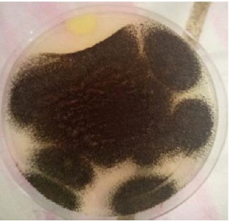
Isolation and identification of fungal isolate: The 1 g of each sample was weighed using an electronic balance and transferred into separate sterile conical flasks containing 100 mL of sterile distilled water each. The samples were then shaken vigorously and an aliquot of 1mL of the mixture was transferred into a sterile test tube containing 9 mL of sterile distilled water using a micropipette and mixed thoroughly, a ten-fold serial dilution of each sample was prepared up to 10-5. Potato Dextrose Agar (PDA) was prepared according to the manufacturer’s protocol and sterilized at 121°C for 15 min in an autoclave Ls-50HD (Wincom™, China). The sterilized media was allowed to cool in a water bath at 45°C and chloramphenicol (50 mg) was added to the media and mixed thoroughly to inhibit bacterial growth. After this, the cooled molten media was dispensed into sterile Petri dishes. An aliquot of 0.1 mL from each dilution was inoculated using the spread plate technique onto the sterile PDA plates. The inoculated plates were then incubated at 28°C for 72 hrs.
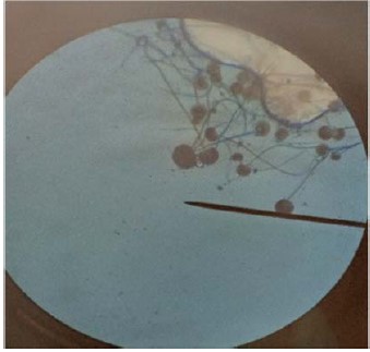
Pure fungal isolates were examined macroscopically and viewed microscopically to characterize their respective spores (Table 1). The macroscopic examination was done by observing the color and texture of the fungal spores on the PDA plates. The microscopic assessment was done using a Motic™ 2820 LED cordless microscope (Wetzlar, Germany), on a Lacto-phenol Cotton Blue (LPCB) mount as described by Cheesbrough (2005)10. The microscopic identification of the fungal spores was confirmed using a Fungi Atlas11. The pure fungi isolate recovered from degraded wood samples were maintained on PDA slants, respectively.
Assessment of fungal isolates for pectinase production: The recovered fungal isolates were kept fresh by repeated transfers on PDA slants. Young cultures were subcultured from fungal stock maintained on slants and incubated at 28°C for 5 days before inoculating their spores into the basal medium. The basal media for assaying the production of pectinase was optimized following the method of Phutela et al.12. The basal medium contained the following constituents in (g/L); peptone, KH2P04 (potassium hydrogen phosphate) 1.05 g, NH4N03 (Ammonium nitrate) 4.0 g, MgS04 7H20 (Magnesium sulfate) 0.5 g, Na2HP04 (Sodium di hydrogen phosphate) 2.0 g and sucrose 20 g12.
The 100 mL of the basal medium was dispensed into eight 250 mL Erlenmeyer flasks, the mouth of the flasks was plugged with non-absorbent cotton wool and wrapped with aluminum foil, respectively. The conical flasks were autoclaved at 121°C for 15 min and allowed to cool to room temperature.
| Table 1: | Macroscopic and microscopic characteristics of the recovered fungal isolates | |||
| Macroscopy | Microscopy (morphology) | Putative fungal isolate |
| Large black colonies | Septate hyphae, non-septate conidiophore, ovoid shape and mop-like conidial head |
Aspergillus niger |
| Yellowish-green, dusty colonies | Septate hyphae, non-septate-conidiophore and columnar-conidial head |
Aspergillus flavus |
| Dark green to grey colonies | Septate hyphae, non-septate conidiophore which was narrow at the base and broad near the vesicle |
Aspergillus fumigatus |
| White fluffy colonies | Non-septate sickle-shaped multicellular macroconidia | Fusarium oxysporum |

|
|
|
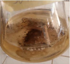
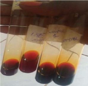
After this, the fungal spores from selected fungal isolates were suspended in 1mL sterile distilled water and aseptically inoculated into each basal medium flask, respectively. A negative control flask was also set up for each of the isolates, but in this case, the medium was not inoculated with any fungal spore. All the flasks were labelled, vigorously shaken at intervals and kept at 30°C for 96 hrs. The flasks were then withdrawn from incubation and assayed for fungal growth and reducing sugar (Fig. 1-2).
The basal media were filtered through a pre-weighed Whatman filter paper and the residues which are mainly mycelial cells, were dried in an oven at 105°C for 3 hrs after which the dry weights were recorded as weight per ml of the sample. The supernatant of the basal media was analyzed for reducing sugar using dinitrosalicyclic acid (DNS) solution. The assay mixture consisted of 3 mls of the supernatant of the basal media and 1mL of sodium acetate buffer was added to each test tube, respectively. The mixtures were incubated at 30°C for 10 min and terminated by the addition of 2 mL DNS solution. The mixtures were heated at 100°C for 5 min and cooled to room temperature. One milliliter (1 mL) of sterile distilled water was added to each of the test tubes and shaken vigorously (Fig. 3). The concentration of reducing sugars in the mixtures was determined using a UV-visible spectrophotometer at 540 nm wavelength (T60 UV-visible spectrophotometer; PG Instrument™, UK).
RESULTS
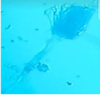
Isolation and identification of fungal isolates: A total of 4 fungal isolates were recovered from the degraded woody samples collected, at the Federal University Dutsin-Ma Take-Off Campus in this study (Fig. 4-6).
The four fungal species recovered include; Aspergillus niger, Aspergillus flavus, Aspergillus fumigatus and Fusarium oxysporum (Table 1).
Among the four fungal species isolated, Aspergillus niger had the highest frequency of occurrence (50%), Aspergillus flavus (25%), Aspergillus fumigatus (15%) and Fusarium oxysporum (10%), respectively (Table 2).
Aspergillus niger had the highest weight per mL sample and Aspergillus fumigatus produced the highest concentration of reducing sugar (0.05 g/mL) among the 3 species of Aspergillus assayed for the production of pectinase in this study (Table 3).
| Table 2: | Frequency of occurrence of fungal species recovered from degraded wood samples | |||
| Fungal species recovered | Number of fungi recovered |
Percentage occurrence of fungi (%) |
| Aspergillus niger | 10 |
50 |
| Aspergillus flavus | 5 |
25 |
| Aspergillus fumigatus | 3 |
15 |
| Fusarium oxysporum | 2 |
10 |
| Total 4 | 20 |
100 |
|
|
|
| Table 3: | Dry weight of fungal hyphae and the concentration of reducing sugar in basal medium | |||
| Fungal isolate | Weight per mL sample |
Absorbance (540 nm) |
Reducing sugar concentration (g/mL) |
| Aspergillus niger | 0.008 |
1.448 |
0.02 |
| Aspergillus flavus | 0.005 |
1.364 |
0.01 |
| Aspergillus fumigatus | 0.006 |
1.345 |
0.05 |
DISCUSSION
In this study, the isolation of 4 species of fungi recovered from degraded woody samples was reported (Table 1). Aspergillus niger had the highest frequency of occurrence (50%) and Fusarium oxysporum had the lowest frequency of occurrence (10%) among the recovered fungal isolates (Table 2). The recovered fungal species are widely distributed and abundant in nature. They are also present on termites and are responsible for soft rot attacks in wood. This was consistent with the reports of Abdel-Azeem et al.13, who reported that Aspergillus niger, Aspergillus fumigatus, Aspergillus flavus and Fusarium oxysporum are among the fungal isolates responsible for soft rot attacks in sections of wood remains in a large archaeological site located in Abydos, in upper Egypt. They also reported that termites’ attack on wooden objects predisposes them to fungal attacks13. Thus, there is a need for further studies to elucidate the relationship between termites and fungi during wood degradation13.
Aspergillus fumigatus produced the highest concentration of reducing sugar (0.05 g/mL) among the 3 species of Aspergillus assayed for the production of pectinase in this study (Table 3).
This infers that Aspergillus fumigatus produced higher carbohydrate-degradative enzymes in the basal medium and may be more vicious in degrading wood during an attack. The enzyme pectinase is used for degrading wood and wooden products by the fungal species. Aspergillus and Penicillium pectinases are commercially valuable products14-15 and are useful in reducing bitterness and cloudiness in fruit juices, as well as degumming fiber crops and improving paper quality14,16.
Basidiomycetes compete in wood degradation by outcompeting their rivals for resources. They do this by increasing their pectinase activity, which facilitates and accelerates the decomposition process and allows them to colonize the wood more rapidly. By secreting higher levels of early-stage colonization enzymes, such as pectinases and oxidant-producing enzymes, the fungi can capture resources at a faster rate than their competitors17-22. Both plants and microorganisms produce pectinolytic enzymes, with plant pectinases aiding in fruit maturation and softening. However, microorganism-produced pectinase breaks down plant matter for use in soil fertilizer and nutrient recycling12,23,24. Thus, this strain of Aspergillus fumigatus can be harnessed for the industrial production of the enzyme pectinase for commercial purposes through genetic engineering and biotechnology25,26.
The Aspergillus niger recovered in the current study had the highest dry weight per mL of fungal mycelium and hyphae (0.008 g/mL) of all the dry weights recorded. This suggested that Aspergillus niger hyphae grow and penetrate deeper into the wood they attack. Fungi are capable of exerting enough mechanical force to push their hyphae through metal foil without the need for enzymes or catalysts4. Fungi that degrade wood initially spread through the woody materials by germinating spores or mycelial fragments, forming hyphae that grow end-to-end. These hyphae seek out interconnecting cell wall pits to extend through the wood and form a mycelial mat. During this initial growth phase, the fungi consume stored products in the parenchyma for energy and build biomass. As decay progresses, the fungi secrete enzymes and metabolites to depolymerize and digest invaded polymeric components of wood, resulting in mass loss that can be measured in laboratory studies4.
Thus, understanding how fungi affect woody materials is imperative and could help to take advantage of current wood coatings to devise strategies that prevent the attack on both the coating materials and the wood they are supposed to safeguard4.
The study highlighted the significance of identifying the fungi responsible for wood decay and underscored the need for effective preservation strategies to prevent wood and wooden materials decay in natural ecosystems.
The research findings can be used to develop effective preservation strategies for wood and wooden materials in the study locality and can open up new ideas for further research in this field. The study findings can serve as a baseline for future research on wood decay fungi in other locations and provide a starting point.
Future studies on wood-decaying fungi should be expanded to include a larger sample size from various locations to understand their prevalence better.
The sample size used in the study was relatively small, which limits the generalizability of the results to other locations within the state. Further studies are required to molecularly characterize the fungal isolates to facilitate comparison with other similar studies.
CONCLUSION
This study has identified four different types of fungi from degraded woody samples, with Aspergillus niger being the most common. Aspergillus fumigatus produced the highest reducing sugar concentration in the optimized basal medium among the three species of Aspergillus tested. The study suggests that Basidiomycetes can outcompete other fungi for resources by increasing pectinase activity. The research highlights the importance of understanding how fungi affect woody materials to develop strategies to prevent attacks on coating materials and wood. Overall, the study provided insights into the degradation process of wood by fungi in the study locality and may provide new ideas and directions for future investigations in this particular field.
SIGNIFICANCE STATEMENT
The deterioration of wood by wood-degrading fungi constitutes a significant source of economic losses. This study was conducted to isolate and characterize fungi responsible for the degradation of wood and determine their pectinolytic activity. The results showed that Ascomycetes and Basidiomycetes were the predominant fungi, with Aspergillus niger being the most frequently occurring species. In addition, Aspergillus fumigatus produced the highest concentration of reducing sugar in the basal medium. These findings provided insights into the types of fungi responsible for wood deterioration in this region and their pectinolytic activities. This information can help in developing effective preservation strategies for wood and wooden materials in the study locality and beyond.
REFERENCES
- Butterfield, B., 2006. The Structure of Wood: Form and Function. In: Primary Wood Processing: Principles and Practice, Walker, J.C.F. (Ed.), Springer, Dordrecht, Netherlands, ISBN: 978-1-4020-4393-2, pp: 1-22.
- Chagnon, C., G. Moreau, C. Bombardier-Cauffopé, J. Barrette, F. Havreljuk and A. Achim, 2022. Broad-scale wood degradation dynamics in the face of climate change: A meta-analysis. GCB Bioenergy, 14: 941-958.
- Cragg, S.M., G.T. Beckham, N.C. Bruce, T.D.H. Bugg and D.L. Distel et al., 2015. Lignocellulose degradation mechanisms across the tree of life. Curr. Opin. Chem. Biol., 29: 108-119.
- Goodell, B., J.E. Winandy and J.J. Morrell, 2020. Fungal degradation of wood: Emerging data, new insights and changing perceptions. Coatings, 10.
- Hamed, S.A.M., 2013. In-vitro studies on wood degradation in soil by soft-rot fungi: Aspergillus niger and Penicillium chrysogenum. Int. Biodeterior. Biodegrad., 78: 98-102.
- Kantharaj, P., B. Boobalan, S. Sooriamuthu and R. Mani, 2017. Lignocellulose degrading enzymes from fungi and their industrial applications. Int. J. Curr. Res. Rev., 9: 1-12.
- Fukasawa, Y., 2021. Ecological impacts of fungal wood decay types: A review of current knowledge and future research directions. Ecol. Res., 36: 910-931.
- Nilsson, T., 2009. Biological Wood Degradation. In: Wood Chemistry and Wood Biotechnology, Ek, M., G. Gellerstedt and G. Henriksson (Eds.), de Gruyter, Berlin, Germany, ISBN: 9783110213409, pp: 219-244.
- Abdulmumini, L., E.L. Irmiya and I. Musab, 2023. Factors influencing youth participation in maize production in Dutsin-MA Local Government Area, Katsina State. J. Agric. Ext., 28: 59-67.
- Cheesbrough, M., 2005. District Laboratory Practice in Tropical Countries, Part 2. Cambridge University Press, England, ISBN: 9780521676311, Pages: 440.
- Levetin, E., 2004. An atlas of fungal spores. J. Allergy Clin. Immunol., 113: 366-368.
- Phutela, U., V. Dhuna, S. Sandhu and B.S. Chadha, 2005. Pectinase and polygalacturonase production by a thermophilic Aspergillus fumigatus isolated from decomposting orange peels. Braz. J. Microbiol., 36: 63-69.
- Abdel-Azeem, A.M., B.W. Held, J.E. Richards, S.L. Davis and R.A. Blanchette, 2019. Assessment of biodegradation in ancient archaeological wood from the Middle Cemetery at Abydos, Egypt. PLoS ONE, 14.
- Silva, D., K. Tokuioshi, E. da Silva Martins, R. da Silva and E. Gomes, 2005. Production of pectinase by solid-state fermentation with Penicillium viridicatum RFC3. Process Biochem., 40: 2885-2889.
- Botella, C., A. Diaz, I. de Ory, C. Webb and A. Blandino, 2007. Xylanase and pectinase production by Aspergillus awamori on grape pomace in solid state fermentation. Process Biochem., 42: 98-101.
- Patidar, M.K., S. Nighojkar, A. Kumar and A. Nighojkar, 2018. Pectinolytic enzymes-solid state fermentation, assay methods and applications in fruit juice industries: A review. 3 Biotech, 8.
- Boddy, L., 2000. Interspecific combative interactions between wood-decaying basidiomycetes. FEMS Microbiol. Ecol., 31: 185-194.
- Hiscox, J., M. Savoury, I.P. Vaughan, C.T. Müller and L. Boddy, 2015. Antagonistic fungal interactions influence carbon dioxide evolution from decomposing wood. Fungal Ecol., 14: 24-32.
- Hori, C., J. Gaskell, K. Igarashi, P. Kersten, M. Mozuch, M. Samejima and D. Cullen, 2014. Temporal alterations in the secretome of the selective ligninolytic fungus Ceriporiopsis subvermispora during growth on aspen wood reveal this organism's strategy for degrading lignocellulose. Appl. Environ. Microbiol., 80: 2062-2070.
- Boddy, L. and J. Hiscox, 2016. Fungal ecology: Principles and mechanisms of colonization and competition by saprotrophic fungi. Microbiol. Spectr., 4.
- Zhang, J., G.N. Presley, K.E. Hammel, J.S. Ryu and J.R. Menke et al., 2016. Localizing gene regulation reveals a staggered wood decay mechanism for the brown rot fungus Postia placenta. Proc. Natl. Acad. Sci. USA, 113: 10968-10973.
- Zhang, J., K.A.T. Silverstein, J.D. Castaño, M. Figueroa and J.S. Schilling, 2019. Gene regulation shifts shed light on fungal adaption in plant biomass decomposers. mBio, 10.
- Suhaimi, H., D.J. Dailin, R. Abd Malek, S.Z. Hanapi and K.K. Ambehabati et al., 2021. Fungal Pectinases: Production and Applications in Food Industries. In: Fungi in Sustainable Food Production, Dai, X., M. Sharma and J. Chen (Eds.), Springer, Cham, New York City, ISBN: 978-3-030-64406-2, Pages: 85-115.
- Palomäki, T. and H.T. Saarilahti, 1997. Isolation and characterization of new C-terminal substitution mutations affecting secretion of polygalacturonase in Erwinia carotovora ssp. carotovora. FEBS Lett., 400: 122-126.
- Okonji, R.E., B.O. Itakorode, J.O. Ovumedia and O.S. Adedeji, 2019. Purification and biochemical characterization of pectinase produced by Aspergillus fumigatus isolated from soil of decomposing plant materials. J. Appl. Biol. Biotechnol., 7: 1-8.
- Anisa, S.K., S. Ashwini and K. Girish, 2013. Isolation and screening of Aspergillus spp. for pectinolytic activity. Electron. J. Biol., 9: 37-41.
How to Cite this paper?
APA-7 Style
Alabi,
E.D., Kilani,
A.M. (2024). Characterization of Wood Degrading Fungi and Their Pectinase Activities in Federal University Dutsin-Ma, Katsina State-Nigeria. Asian Science Bulletin, 2(2), 174-182. https://doi.org/10.3923/asb.2024.174.182
ACS Style
Alabi,
E.D.; Kilani,
A.M. Characterization of Wood Degrading Fungi and Their Pectinase Activities in Federal University Dutsin-Ma, Katsina State-Nigeria. Asian Sci. Bul 2024, 2, 174-182. https://doi.org/10.3923/asb.2024.174.182
AMA Style
Alabi
ED, Kilani
AM. Characterization of Wood Degrading Fungi and Their Pectinase Activities in Federal University Dutsin-Ma, Katsina State-Nigeria. Asian Science Bulletin. 2024; 2(2): 174-182. https://doi.org/10.3923/asb.2024.174.182
Chicago/Turabian Style
Alabi, Emmanuel, Dayo, and Adetunji Misbau Kilani.
2024. "Characterization of Wood Degrading Fungi and Their Pectinase Activities in Federal University Dutsin-Ma, Katsina State-Nigeria" Asian Science Bulletin 2, no. 2: 174-182. https://doi.org/10.3923/asb.2024.174.182

This work is licensed under a Creative Commons Attribution 4.0 International License.